Canadian experts release first molecular images of B.1.1.7 variant of Covid-19
Canadian experts release first molecular images of B.1.1.7 variant of Covid-19on May 04, 2021
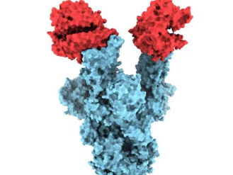
B.1.1.7 variant of Covid-19
Canadian experts
Coronavirus
Covid-19
Frontlist
Frontlist India
Frontlist Latest
Frontlist Latest news
Frontlist Latest Update
Frontlist News
release first molecular image
Post a comment
0 comments

Trending Posts
.jpg)
.jpg)


.jpg)
_(1).jpg)
.jpg)

.jpg)
Recent Post

.jpg)
.jpg)
.jpg)
.jpg)

.jpg)
Related Post


© 2026 - Frontlist - News and Updates of Publishing Industry, Book Reviews, Education News, Author Interviews, and Videos.. All Rights Reserved.
Sorry! No comment found for this post.